Geniculate ganglion
Collection of facial nerve neurons
| Geniculate ganglion | |
|---|---|
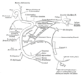
 The course and connections of the facial nerve in the temporal bone. | |
 Cranial nerves VII and VIII and selected structures of the inner and middle ear. 1 Nervus vestibularis, 2 Nervus cochlearis, 3 Nervus intermediofacialis, 4 Ganglion geniculi, 5 Chorda tympani, 6 Cochlea, 7 Ductus semicirculares, 8 Malleus, 9 Membrana tympani, 10 Tuba auditiva | |
| Details | |
| Innervates | Lacrimal glands, submandibular glands, sublingual glands, tongue, palate, pharynx, external auditory meatus, stapedius muscle, posterior belly of the digastric muscle, stylohyoid muscle, muscles of facial expression |
| Identifiers | |
| Latin | ganglion geniculi nervi facialis |
| MeSH | D005830 |
| TA98 | A14.2.01.116 |
| TA2 | 6287 |
| FMA | 53414 |
| Anatomical terms of neuroanatomy [edit on Wikidata] | |
The geniculate ganglion (from Latin genu, for "knee"[1]) is a bilaterally paired special sense ganglion[2] of the intermediate nerve component of the facial nerve (CN VII).[3] It is situated within facial canal of the head.[citation needed]
It contains cell bodies of first-order unipolar sensory neurons which convey gustatory (taste) afferents from taste receptors of the anterior two-thirds of the tongue by way of the chorda tympani, and of the palate by way of the greater petrosal nerve, From the gangion, the proximal fibres proceed to the gustatory (i.e. superior/rostral[3]) part of the solitary nucleus where they synapse with second-order neurons.[2]
Anatomy
Structure
The geniculate ganglion is conical in shape. The greater petrosal nerve diverges from CNVII and the lesser petrosal nerve diverges from CN IX at the geniculate ganglion.[3]
Relations
It is located close to the internal auditory meatus.[4] It is covered superiorly by the petrous part of the temporal bone (which is sometimes absent over the ganglion).[5]
Clinical significance
The geniculate ganglion is an important surgical landmark near the internal auditory meatus.[4]
Herpes zoster oticus
The geniculate ganglion may become inflamed due herpes zoster virus virus infection. Swelling of the ganglion may result in facial palsy (Ramsay Hunt syndrome). The syndrome presents with intense pain in one ear that is followed by a vesicular rash around the ear canal.[2]
Additional images
-
 Plan of the facial and intermediate nerves and their communication with other nerves.

See also
References
- ^ "genu-, geni-, gen- + (Latin: knee)". WordInfo. Retrieved 2008-10-03.
- ^ a b c Fitzgerald, Maurice J. T.; Gruener, Gregory; Mtui, Estomih (2011). Clinical Neuroanatomy and Neuroscience (6th ed.). Edinburgh: Saunders. pp. 241–243. ISBN 978-0-7020-3738-2.
- ^ a b c "ganglion géniculé l.m. - Dictionnaire médical de l'Académie de Médecine". www.academie-medecine.fr. Retrieved 2024-05-24.
- ^ a b Hall, George M.; Pulec, Jack L.; Rhoton, Albert L. Jr. (1 November 1969). "Geniculate Ganglion Anatomy for the Otologist". Archives of Otolaryngology. 90 (5): 568–571. doi:10.1001/archotol.1969.00770030570007. ISSN 0003-9977.
- ^ Rhoton, Albert L.; Pulec, Jack L.; Hall, George M.; Boyd, Allen S. (1 January 1968). "Absence of Bone over the Geniculate Ganglion". Journal of Neurosurgery. 28 (1): 48–53. doi:10.3171/jns.1968.28.1.0048.
External links
- cranialnerves at The Anatomy Lesson by Wesley Norman (Georgetown University) (VII)
- lesson3 at The Anatomy Lesson by Wesley Norman (Georgetown University) (midearcavity)
- v
- t
- e
The cranial nerves
- Nuclei
- Course
- no significant branches
- Nuclei
- Course
- Nuclei
- Course
- Nuclei
- Branches
- Nucleus
- Branches
- no significant branches
- Nuclei
- Course
- Branches
- Nucleus
- Branches
- no significant branches
| Near origin |
|
|---|---|
| Inside facial canal | |
| At stylomastoid foramen | |
| Nuclei |
| Before jugular fossa | |
|---|---|
| After jugular fossa | |
| Nuclei |
| Before jugular fossa | |
|---|---|
| After jugular fossa | |
| Neck | |
| Thorax |
|
| Abdomen | |
| Nuclei |
| Authority control databases |
|
|---|


