BTLA
Protein-coding gene in the species Homo sapiens
| BTLA | |||||||||||||||||||||||||||||||||||||||||||||||||||
|---|---|---|---|---|---|---|---|---|---|---|---|---|---|---|---|---|---|---|---|---|---|---|---|---|---|---|---|---|---|---|---|---|---|---|---|---|---|---|---|---|---|---|---|---|---|---|---|---|---|---|---|
 | |||||||||||||||||||||||||||||||||||||||||||||||||||
| Identifiers | |||||||||||||||||||||||||||||||||||||||||||||||||||
| Aliases | BTLA, BTLA1, CD272, B and T lymphocyte associated | ||||||||||||||||||||||||||||||||||||||||||||||||||
| External IDs | OMIM: 607925; MGI: 2658978; HomoloGene: 52233; GeneCards: BTLA; OMA:BTLA - orthologs | ||||||||||||||||||||||||||||||||||||||||||||||||||
| |||||||||||||||||||||||||||||||||||||||||||||||||||
| |||||||||||||||||||||||||||||||||||||||||||||||||||
| |||||||||||||||||||||||||||||||||||||||||||||||||||
| |||||||||||||||||||||||||||||||||||||||||||||||||||
| |||||||||||||||||||||||||||||||||||||||||||||||||||
| Wikidata | |||||||||||||||||||||||||||||||||||||||||||||||||||
| |||||||||||||||||||||||||||||||||||||||||||||||||||


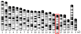

B- and T-lymphocyte attenuator or BTLA (also known as cluster of differentiation 272 or CD272) is a protein that belongs to the CD28 immunoglobulin superfamily (IgSF) which is encoded by the BTLA gene located on the 3rd human chromosome.[5][6] BTLA was first discovered in 2003 as an inhibitor of Th1 expansion and it became the 3rd member of the CD28 IgSF. However, its discovered ligand herpes virus entry mediator or HVEM (also known as tumour necrosis factor receptor superfamily member 14 or TNFRSF14) belongs to the tumor necrosis factor receptor superfamily (TNFRSF). This finding was surprising because until the discovery of HVEM it was believed that receptors and ligands always belong to the same family.[7][8]
Expression
BTLA is broadly expressed in various organs. Among these are the lymph nodes, the thymus and the spleen where high expression of BTLA can be found. On the contrary, low to no expression is detected in organs such as the liver, kidney, heart, brain and other organs. If we consider specific cell populations then the expression of BTLA can be ascertained in T cell, B cells, DCs and NKT cells. In most of the aforementioned cells BTLA expression fluctuates depending on different developmental stages. For instance, in T cells, in which BTLA is most frequently studied, BTLA is stably expressed in naive T cells, the expression subsequently increases when a T cell is stimulated, but once fully activated the expression of BTLA again decreases.[7][9]
Structure
BTLA is a 289 amino acid long transmembrane glycoprotein. It is very similar in structure to PD-1 and CTLA-4. Therefore, it consists of an extracellular domain, a transmembrane domain and a cytoplasmic domain. The cytoplasmatic domain is indispensable for signalling and it is constituted of 3 important motives: the growth factor receptor-bound protein-2 (Grb-2) recognition motif, the immunoreceptor tyrosine-based inhibitory motif (ITIM) and the immunoreceptor tyrosine-based switch motif (ITSM).[7]
Function
Considering the above-mentioned motives in the cytoplasmatic part of BTLA, it is obvious that BTLA has inhibition and activation capacities. The reason is that through ITIM it recruits either SHP-1 or SHP-2 which act as phosphatases and dephosphorylate tyrosine which suppresses immune activation. On the other hand, it also possesses the Grb-2 recognition motif which, when recognized by a Grb-2 protein, promotes activation of PI3K and that further mediates cell proliferation and survival.[7][9]
As stated above, HVEM is the main ligand of BTLA but unlike BTLA, HVEM is able to interact with other molecules as well namely CD160 and tumor necrosis factor superfamily member 14 (TNFSF14) also known as LIGHT. Interaction of HVEM and CD160 is inhibitory but interaction with LIGHT is stimulatory. Therefore, HVEM can also induce both stimulation and inhibition. Taken together, it seems that the molecules CD160, BTLA, LIGHT and HVEM form a kind of regulatory network.[7][8][9]
T cell
Both BTLA and HVEM are expressed on naive T cells. This colocalization on a single cell allows them to interact in a cis manner (on the same cell). This interaction is important in immunological tolerance as shown by the fact that BTLA mouse knockdown is unable to develop tolerance to high doses of ovalbumin.[7] Furthermore, the knockdown also proves that BTLA is dispensable for T cell development as no abnormalities were detected in T cells development.[9]
When a T cell is fully activated HVEM is internalized and this allows BTLA to interact in a trans manner (with molecules on other cells).[9] This enables other cells who express HVEM to regulate activated T cells. Treg cells express HVEM and are able to facilitate immune suppression through interaction with BTLA.[7] Furthermore, the blockage of BTLA together with PD-1 can help reverting the state of exhaustion which further proves the importance of BTLA as an inhibitory molecule.[9] However, in some cases interaction of BTLA and HVEM can have an opposite effect. For instance, in the case of infection with vaccinia virus BTLA and HVEM interaction promotes survival of CD8+ T cells and production of memory cells.[7]
B cell
B cell's expression of BTLA is less studied than T cell's but it is clear that peripheral B cells express a high amount of BTLA whereas in bone marrow B cell precursors express a lower amount of BTLA. BTLA has been connected with suppressory function in B cells and its expression is not necessary for B cell development just like in T cells. However, a lower expression of BTLA on B cells in elderly is implied in a less potent response to the influenza vaccine.[7][9]
DC
BTLA can also be found on DCs, with mature DCs having a higher expression of BTLA and immature DCs a lower one. Just like in T cells and B cells, most results show that BTLA and HVEM interaction suppresses the maturation of DCs.[7][9]
Clinical significance
Tumors
In many cases BTLA expression is connected with unfavourable outcomes as it, for instance, inhibits the function of human CD8+ cancer-specific T cells.[10] For example, this is the case of gallbladder cancer where BTLA+ CD8+ T cells are associated with inhibition of anticancer immunity. Similarly, BTLA+ CD8+ T cells show partial dysfunctionality and decrease secretion of IFN-γ in melanoma patients. Therefore, it seems that BTLA can be used as a novel antitumor therapy target. This notion is supported by the fact that in patients with hepatocarcinoma blocking of BTLA signalling increases the secretion of IFN-γ by CD8+ and CD4+ T cells.[9] However, some studies have shown that, for instance, colorectal carcinoma is associated with lower BTLA expression and that the lower BTLA expression is connected with poor survival.[11] Even in some cases of melanoma BTLA+ CD8+ lymphocytes are suggested to be connected with better outcomes of adoptive cell therapy. Hence, it seems that BTLA has rather a context-specific function. This fact should be taken into account if BTLA is to be used as a cancer therapy target.[9]
Infectious diseases
As in the case of tumors, BTLA seems to be mainly an inhibitory molecule as, for instance, in the case of pulmonary tuberculosis where BTLA expression in CD4+ and CD8+ T cells is connected with the disease progression.[9] Similar results can be seen during helminth infections. When mice are infected by Strongyloides ratti the mice with BTLA knockdown are faster at eliminating the parasite from the intestine probably through the production of IL-9.[12] Furthermore, the course of virus infections seems to be negatively affected by BTLA as well. During chronic hepatitis B infection BTLA impairs T cell response and what's more blocking BTLA leads to T cell proliferation and higher cytokine production. However, as already mentioned in some cases BTLA seems to have a stimulatory role. This is the case of the aforementioned pulmonary tuberculosis where BTLA+ αβ T cells are predominantly of central memory phenotype and fight off Mycobacterium tuberculosis. A further example is an infection with vaccinia virus during which deletion of BTLA or HVEM causes instability and early apoptosis of CD8+ T cells.[9]
References
- ^ a b c GRCh38: Ensembl release 89: ENSG00000186265 – Ensembl, May 2017
- ^ a b c GRCm38: Ensembl release 89: ENSMUSG00000052013 – Ensembl, May 2017
- ^ "Human PubMed Reference:". National Center for Biotechnology Information, U.S. National Library of Medicine.
- ^ "Mouse PubMed Reference:". National Center for Biotechnology Information, U.S. National Library of Medicine.
- ^ "Entrez Gene: BTLA B and T lymphocyte associated".
- ^ Watanabe N, Gavrieli M, Sedy JR, Yang J, Fallarino F, Loftin SK, et al. (July 2003). "BTLA is a lymphocyte inhibitory receptor with similarities to CTLA-4 and PD-1". Nature Immunology. 4 (7): 670–9. doi:10.1038/ni944. PMID 12796776. S2CID 11943145.
- ^ a b c d e f g h i j Yu X, Zheng Y, Mao R, Su Z, Zhang J (2019). "BTLA/HVEM Signaling: Milestones in Research and Role in Chronic Hepatitis B Virus Infection". Frontiers in Immunology. 10: 617. doi:10.3389/fimmu.2019.00617. PMC 6449624. PMID 30984188.
- ^ a b Pasero C, Olive D (March 2013). "Interfering with coinhibitory molecules: BTLA/HVEM as new targets to enhance anti-tumor immunity". Immunology Letters. 151 (1–2): 71–5. doi:10.1016/j.imlet.2013.01.008. PMID 23439006.
- ^ a b c d e f g h i j k l Ning Z, Liu K, Xiong H (2021-03-29). "Roles of BTLA in Immunity and Immune Disorders". Frontiers in Immunology. 12: 654960. doi:10.3389/fimmu.2021.654960. PMC 8043046. PMID 33859648.
- ^ Derré L, Rivals JP, Jandus C, Pastor S, Rimoldi D, Romero P, et al. (January 2010). "BTLA mediates inhibition of human tumor-specific CD8+ T cells that can be partially reversed by vaccination". The Journal of Clinical Investigation. 120 (1): 157–67. doi:10.1172/JCI40070. PMC 2799219. PMID 20038811.| *Lay summary in: "Researchers Identify Protein that Inhibits Tumor-Targeting Immune Cells When Activated". Genetic Engineering & Biotechnology News. December 29, 2009.
- ^ Song J, Wu L (2020). "Friend or Foe: Prognostic and Immunotherapy Roles of BTLA in Colorectal Cancer". Frontiers in Molecular Biosciences. 7: 148. doi:10.3389/fmolb.2020.00148. PMC 7385242. PMID 32793631.
- ^ Breloer M, Hartmann W, Blankenhaus B, Eschbach ML, Pfeffer K, Jacobs T (February 2015). "Cutting Edge: the BTLA-HVEM regulatory pathway interferes with protective immunity to intestinal Helminth infection". Journal of Immunology. 194 (4): 1413–6. doi:10.4049/jimmunol.1402510. PMID 25595777.
Further reading
- Pao LI, Bedzyk WD, Persin C, Cambier JC (February 1997). "Molecular targets of CD45 in B cell antigen receptor signal transduction". Journal of Immunology. 158 (3): 1116–24. doi:10.4049/jimmunol.158.3.1116. PMID 9013950. S2CID 24366449.
- Pao LI, Cambier JC (March 1997). "Syk, but not Lyn, recruitment to B cell antigen receptor and activation following stimulation of CD45- B cells". Journal of Immunology. 158 (6): 2663–9. doi:10.4049/jimmunol.158.6.2663. PMID 9058799. S2CID 45350654.
- Vilen BJ, Famiglietti SJ, Carbone AM, Kay BK, Cambier JC (July 1997). "B cell antigen receptor desensitization: disruption of receptor coupling to tyrosine kinase activation". Journal of Immunology. 159 (1): 231–43. doi:10.4049/jimmunol.159.1.231. PMC 3931421. PMID 9200459.
- Gavrieli M, Watanabe N, Loftin SK, Murphy TL, Murphy KM (December 2003). "Characterization of phosphotyrosine binding motifs in the cytoplasmic domain of B and T lymphocyte attenuator required for association with protein tyrosine phosphatases SHP-1 and SHP-2". Biochemical and Biophysical Research Communications. 312 (4): 1236–43. doi:10.1016/j.bbrc.2003.11.070. PMID 14652006.
- Sedy JR, Gavrieli M, Potter KG, Hurchla MA, Lindsley RC, Hildner K, et al. (January 2005). "B and T lymphocyte attenuator regulates T cell activation through interaction with herpesvirus entry mediator". Nature Immunology. 6 (1): 90–8. doi:10.1038/ni1144. PMID 15568026. S2CID 23871512.
- Gonzalez LC, Loyet KM, Calemine-Fenaux J, Chauhan V, Wranik B, Ouyang W, Eaton DL (January 2005). "A coreceptor interaction between the CD28 and TNF receptor family members B and T lymphocyte attenuator and herpesvirus entry mediator". Proceedings of the National Academy of Sciences of the United States of America. 102 (4): 1116–21. Bibcode:2005PNAS..102.1116G. doi:10.1073/pnas.0409071102. PMC 544343. PMID 15647361.
- Cheung TC, Humphreys IR, Potter KG, Norris PS, Shumway HM, Tran BR, et al. (September 2005). "Evolutionarily divergent herpesviruses modulate T cell activation by targeting the herpesvirus entry mediator cosignaling pathway". Proceedings of the National Academy of Sciences of the United States of America. 102 (37): 13218–23. Bibcode:2005PNAS..10213218C. doi:10.1073/pnas.0506172102. PMC 1201609. PMID 16131544.
- Compaan DM, Gonzalez LC, Tom I, Loyet KM, Eaton D, Hymowitz SG (November 2005). "Attenuating lymphocyte activity: the crystal structure of the BTLA-HVEM complex". The Journal of Biological Chemistry. 280 (47): 39553–61. doi:10.1074/jbc.M507629200. PMID 16169851.
- Otsuki N, Kamimura Y, Hashiguchi M, Azuma M (June 2006). "Expression and function of the B and T lymphocyte attenuator (BTLA/CD272) on human T cells". Biochemical and Biophysical Research Communications. 344 (4): 1121–7. doi:10.1016/j.bbrc.2006.03.242. PMID 16643847.
- Wang XF, Chen YJ, Wang Q, Ge Y, Dai Q, Yang KF, et al. (February 2007). "Distinct expression and inhibitory function of B and T lymphocyte attenuator on human T cells". Tissue Antigens. 69 (2): 145–53. doi:10.1111/j.1399-0039.2006.00710.x. PMID 17257317.
External links
- BTLA+protein,+human at the U.S. National Library of Medicine Medical Subject Headings (MeSH)
- Human BTLA genome location and BTLA gene details page in the UCSC Genome Browser.
- Overview of all the structural information available in the PDB for UniProt: Q7Z6A9 (B- and T-lymphocyte attenuator) at the PDBe-KB.
This article incorporates text from the United States National Library of Medicine, which is in the public domain.
- v
- t
- e
PDB gallery
-
 2aw2: Crystal structure of the human BTLA-HVEM complex


